
Highlight
Neutron Scattering Study of Water Diffusion on Single-Supported Bilayer Lipid Membranes

IGERT trainee Andrew Miskowiec at the University of Missouri Research Reactor
Achievement/Results
Profs. Haskell Taub and Flemming Hansen and their team members at the University of Missouri and the Technical University of Denmark have successfully used neutron scattering and computer simulations to characterize the random motion of water molecules associated with a bilayer lipid membrane. Their achievement is significant because bilayer lipid membranes form the boundary of virtually all living cells; and an understanding of the structure and motion of the membrane-associated water is essential for explaining how the membrane functions. Because of its sensitivity to hydrogen atoms, neutron scattering is very effective for probing the motion of membrane-associated water. Unfortunately, due to the weak scattering from a single membrane, previous neutron scattering experiments have used samples consisting of stacks of thousands of membranes supported on a solid substrate. But such experiments are difficult to model in computer simulations. Besides the sheer number of membranes, the interactions between them within a stack, and the presence of unknown amounts of water between membranes complicate the interpretation of the neutron spectra.
Address Goals
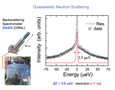
The interdisciplinary team of Taub and Hansen has been able to solve the problem of using stacks of many membranes by depositing a single bilayer membrane onto an oxide-coated silicon substrate as shown in Fig. 1. By stacking a hundred of such wafers, they obtained an adequate intensity of scattered neutrons and yet retained a sample simple enough for simulation on a computer. Using such a sample, they then performed high-energy-resolution neutron scattering experiments at the NIST Center for Neutron Research and the Spallation Neutron Source at Oak Ridge National Laboratory. The neutron scattering spectrometer used in the Oak Ridge measurements is shown in the lower left of Fig. 2. One can appreciate the size of the spectrometer by comparing with Prof. Hansen, who is standing in front of it. This size is necessary to accommodate the many neutron detectors needed to increase the counting rate in the experiment. The long 84-meter flight path shown above the spectrometer enables the energy of the neutrons incident upon a sample to be determined with high precision. Figure 3 shows IGERT trainee, Andrew Miskowiec, performing preliminary neutron scattering measurements at the University of Missouri Research Reactor.
By analyzing the inelastic neutron spectra from water molecules associated with the single-supported membrane sample, the team was able to infer three different types of water motion as indicated in Fig. 1: 1) bulk-like motion of water situated well above the membrane; 2) motion of water confined to a region near the lipid head groups; and 3) motion of bound water that is hydrogen-bonded to the head groups 1. The motion of bulk-like and confined water molecules is fast, compared to those bound to the head groups of the lipid molecules, which move on the same nanosecond time scale as H atoms in the lipid molecules. These results were found to be consistent with computer simulations on a freestanding bilayer lipid membrane 2.
References:
1. M. Bai, A. Miskowiec, F. Y. Hansen, H. Taub, T. Jenkins, M. Tyagi, S. O. Diallo, E. Mamontov, K. W. Herwig, S.-K. Wang, “Study of the Water Diffusion on Single-supported Bilayer Lipid Membranes by Quasielastic Neutron Scattering,” Europhys. Lett. 98, 48006, 1-6 (2012).
2. F. Y. Hansen, G. H. Peters, H. Taub, and A. Miskowiec, “Diffusion of Water and Selected Atoms in DMPC Lipid Bilayer Membranes,” J. Chem. Phys. 137, 204919, 1-15 (2012).