

Highlight Photo
Comparing Brain Activation With Computer Vision Algorithms
Matching Brain Activation Against Computer Models
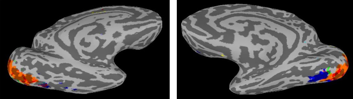
Left and right hemisphere matches of fMRI activity to five vision models: Gabor filterbank (orange), SIFT (blue), geometric blur (green), shock graph (yellow). Separate colors are used for regions matching two models (red) or three or more (purple), including HMAX.
Credits: Daniel Leeds, Carnegie Mellon University
