
Highlight Photo
Actin Interacting Protein 1 and Actin Depolymerizing Factor Drive Rapid Actin Dynamics in Polarized Plant Cells
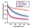
Figure 1. AIP1 and ADF promote in vivo actin dynamics.
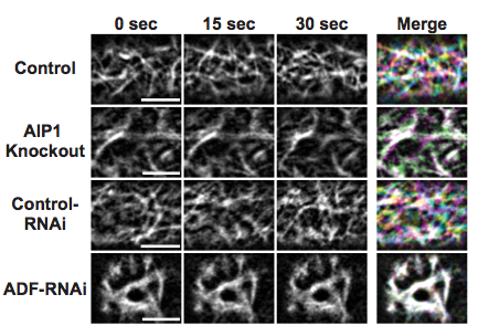
Actin dynamics at the cortex of protonemal cells was visualized by time-lapse confocal microscopy using the actin probe lifeact-mEGFP. Representative images of actin are shown as grayscale images representing cyan, yellow, and magenta for the 0, 15, and 30 second time intervals, respectively. The merge combines all time intervals as separate color channels projected onto one RGB image. White indicates overlap of actin in all time points; color indicates that actin has changed in at least one of the three time points. Scale bar = 5 µm.
Credits: Robert Augustine