

Highlight Photo
The 1.9 structure of human Alpha-N-acetylgalactosaminidase: the molecular basis of Schindler and Kanzaki diseases
Figure 1: Schindler and Kanzaki disease mutations.
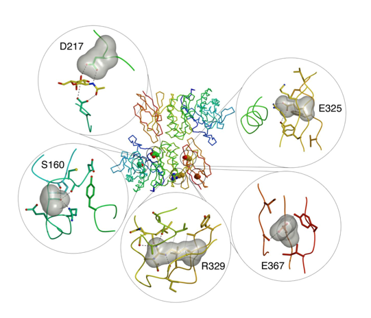
Five residues are shown in the center panel showing the location of the residue in the polypeptide fold. The panels show a surface representation of the ligand as well as the surrounding residues. Hydrogen bonds are shown as black dashed lines and van der Waals interactions as grey lines.
Credits: Clark, N. E., and Garman, S. C.
