
Highlight
Fractal Electrodes for Retinal Implants
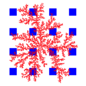
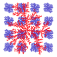
Figure 1: A retinal neuron (red) connecting to 81% of surrounding square electrodes (blue). Underlying photodiodes not shown.
Achievement/Results
There are currently 1.75 million U.S. citizens affected by age-related macular degeneration. This is a degenerative eye disease that affects the rods and cones in the retina, but leaves intact the rest of the retinal circuitry necessary for vision. Research groups around the world have been developing retinal implants to replace the functionality of the eye’s photoreceptors and restore vision to those affected by this and other similar diseases.
A typical retinal implant is a 3mm device consisting of an array of micro-photodiodes that interact with the retinal neurons via electrodes. Due to the peculiar architecture of the human eye, where light must pass through all the retinal circuitry before being absorbed by the photosensitive cells, the electrodes must be positioned in front of the photodiodes in order to communicate with the retinal neurons.
Unfortunately, this setup blocks some of the light from being absorbed by the photodiodes. Another shortcoming of these implants is the resolution lost because of the poor connection between the implant and the retinal neurons. See Figure 1 for a representation of the connection made between the electrodes (blue) on the photodiodes (not shown) and the retinal neuron (red). The neuron is able to connect to only a portion of the surrounding electrodes, specifically 13 of 16 electrodes are connected to the neuron, providing a yield of 81%.
Working in the physics lab of Professor Richard Taylor (University of Oregon), IGERT trainee Rick Montgomery’s research has focused on a solution to these two problems. In a collaborative effort with Professor Simon Brown at the University of Canterbury in Christchurch, NZ, the researchers drew on the Taylor group’s knowledge of fractals and the Brown group’s expertise in nanocluster deposition and aggregate growth to design an electrode that reduces the deficiencies of the retinal implants.
Fractals are objects that are similar to themselves at different magnifications and they exhibit many beneficial properties, such as high boundary-to-surface-area ratios. In particular, the large boundary-to-surface-area ratio is beneficial for reaching far out in space while not occupying a lot of area. Replacing the square electrodes with those of a fractal shape (represented in Figure 2) will increase the number of electrodes that will be in contact with retinal neurons (providing a yield of 94%), while also increasing the amount of light that can pass through the electrodes, due to their branched, fractal structures.
These electrodes are grown by depositing nanoparticles on smooth substrates in ultra-high vacuum chambers. The nanoparticles diffuse across the surface until encountering a nucleation site. As more and more particles are deposited on the substrate, the aggregates grow in size all the while maintaining this branched, fractal structure. Thus the fractal electrodes can be grown to the size of the micro-photodiodes providing that beneficial connectivity while not hindering the photodiode’s light-collecting capabilities.
A patent application for this novel design was filed in February 2011. An IGERT international travel award will allow Montgomery to visit Prof. Brown’s lab in New Zealand in May 2011 to collaborate on future aspects of this research project.
Address Goals
Advances in Human health represent one of great goals of science. In particular, exploiting the rapid advances in nanoelectronics research for human implants has the potential to revolutionize many surgical strategies and herald major improvements across society. Development of retinal implants provides the opportunity to restore vision to hundreds of thousands of people around the world. In addition to applications, investigations of the biological-nanoelectronics interface will lead to advances in fundamental phenomena in biophysics.