
Highlight Photo
Potential Gene Transfer Adjuvants to Improve Intrathecal Microencapsulated Interleukin-10 Gene Delivery
Fluorescent Tagged DNA
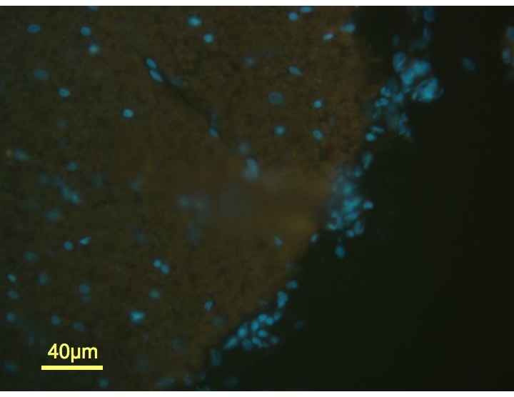
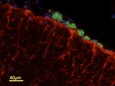
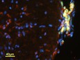
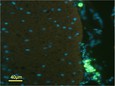
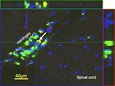
Fig. 1 Fluorescent tagged DNA remains associated with meningeal macrophage cells but not the spinal parenchyma with no evidence of cellular apoptosis. Fluorescent histological examination of spinal cord sections (n=3) near the lumbar spinal cord injection site, segments L3-4, 8 weeks after i.t. injection of DOTAP protocells loaded with FAM-tagged 18bp DNA oligomer. Protocell delivered DNA (green; white arrow) is not colocalizing with astroglia stained for glial fibrillary activating protein, GFAP (A; red) but is co-localizing in the meninges with activated macrophage stained for OX2 (B; red plus green DNA = yellow; white arrow). There is no evidence of cellular death in the meninges or spinal cord as there is no positive staining for the apoptotic marker, activated Caspase 3 (absence of red) in protocell treated (C; green; white arrow) compared to nave animals (D). Cell nuclei are stained with DAPI (blue). All images are at 20X.
Credits: Ellen Dengler, University of New Mexico