
Highlight Photo
Getting in the groove: evidence of chemical communication in the primate fossil record
Figure 2. The vomeronasal groove (VNG) as seen in three modalities
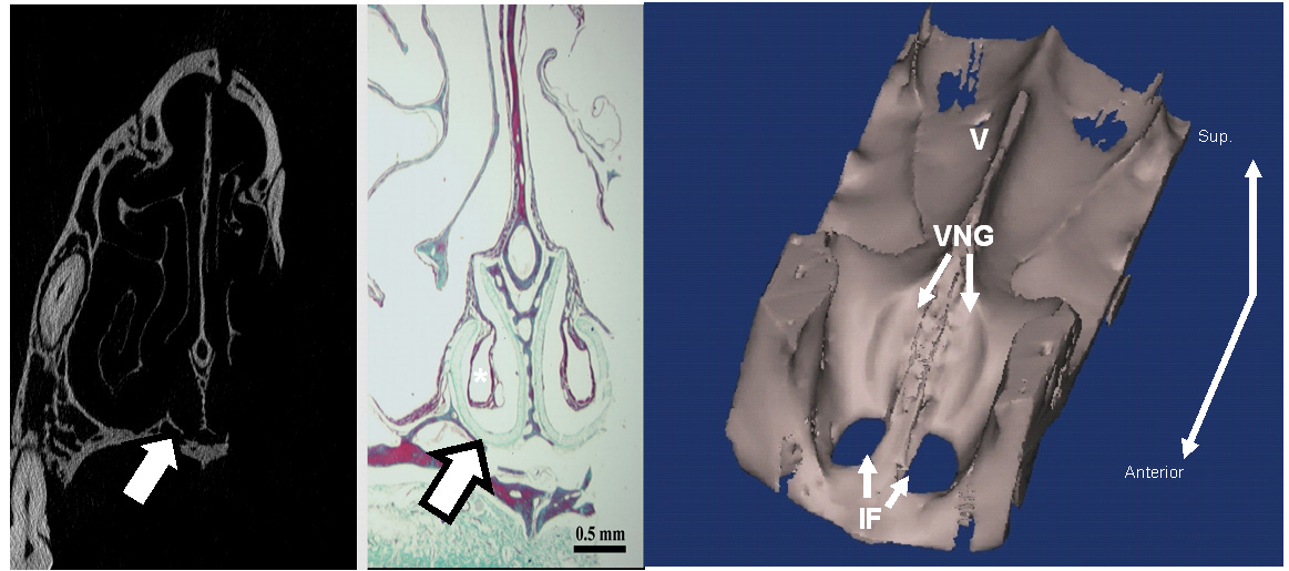
On the left, a CT scan slice across the nasal region of a galago (a lorisoid lower primate) reveals the vomeronasal groove (VNG) as a cup-shaped depression (arrow). In the middle view, from a histological slice through soft tissue of the same animal, the VNG is seen as it relates to surrounding tissues; the white asterisk above the arrow is the opening of the VNO which receives chemical stimuli. On the right, a full CT scan has been rendered to show a reconstruction of the entire floor of the nasal cavity: the upper teeth cannot be seen but are located at the lower left (near) end; behind that are the twin openings of the incisive foramina connecting the nasal cavity with the mouth below; behind and between them is the vomer (V). Alongside the vomer just behind the incisive foramina are the paired vomeronasal grooves (VNG) into which fit the cartilaginous support for the VNO.
Credits: Courtesy of and copyright Eva Garrett