
Highlight
Elucidating the Role of Dynamics in IgE Receptor Signaling via Single Quantum Dot Tracking
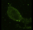
Confocal image series compressed into a single plane, depicting a live RBL-2H3 cell labeled with three spectrally distinct quantum dot-based probes.
Achievement/Results
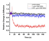
Researchers at the University of New Mexico (UNM) and Sandia National Laboratories (SNL) are probing the mechanisms of allergic inflammation at the level of single molecules. Through funding from the NSF’s Integrated Graduate Education and Research Traineeship program on Integrating Nanotechnology with Cell Biology and Neuroscience, this interdisciplinary project brings together cell biologists, material scientists, organic chemists, engineers, and physicists. The study focuses on the high affinity immunoglobulin E receptor; referred to as FceRI. This receptor is present on histamine-containing cells in the body and, in response to an allergen, initiates a cellular response that includes the release of histamine and other pro-inflammatory molecules into the surrounding environment. This surge of pro-inflammatory molecules produces many of the symptoms associated with seasonal allergies, allergic asthma, and other conditions, such as urticaria and anaphylaxis. While the signaling events that occur after activation of the receptor have been intensely studied for many years, the precise mechanism by which exposure to allergen activates the receptor has remained elusive, due in part to technical limitations associated with imaging of individual molecules in living cells. Collaboration between the laboratories of Prof. Diane Lidke at the UNM School of Medicine and Dr. Timothy Boyle at SNL Advanced Materials Laboratory, has produced novel nanoparticle-based probes enabling the study of FceRI signaling at the level of individual receptors (Figure 1) and allergens (Figure 2). Using these probes in combination with state-of-the-art microscopy methods and biophysical techniques developed through collaboration between Dr. Lidke’s group and physicists and engineers from both UNM and SNL, the dynamic behavior and spatial distribution of individual receptors and allergen-receptor complexes has been characterized in both unstimulated and allergen-exposed cells. The interdisciplinary team has demonstrated that the previously described rapid (<20 s) transition from small, dynamic clusters to large, stable signaling domains in response to multi-valent antigen binding is dependent upon an intact cytoskeleton and also highly dependent on antigen concentration (Figure 3). In fact, at comparatively low antigen doses, at which maximal cellular response occurs, there is relatively little change in FceRI diffusion. These studies challenge the paradigm that FceRI immobilization is a pre-requisite for signal initiation.
Address Goals
The above-described project contributes significantly to our understanding of the allergic inflammatory response through the application of novel, nanoparticle-based live cell imaging probes and cutting-edge microscopy techniques. This research not only provides new insights into FceRI signaling, but also highlights the importance of cell membrane topography in governing signal transduction; an area that has received little attention so far, primarily due to technical limitations. Through the dissemination of these discoveries, it is expected that other researchers will be inspired to examine the roles of receptor diffusion and cell membrane topography in their own particular areas of interest, yielding new insights into a wide variety of cellular phenomena. The development and application of state-of-the-art equipment and image processing algorithms has been central to this research. NSF INCBN IGERT funding has facilitated the purchase of several pieces of high-end microscopy and data storage hardware which has been added to the microscopy facility shared resource at the UNM School of Medicine. By making such equipment available to the wider scientific community at UNM, NSF is promoting the advancement of not only the current research described above, but also providing tools to broaden the research horizons of dozens of scientists at the University of New Mexico.